[最新] microscope e coli morphology 308954-E coli morphology under microscope
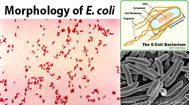
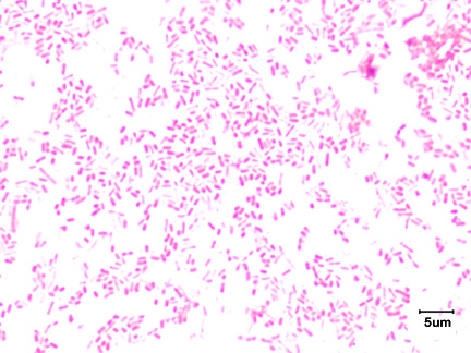
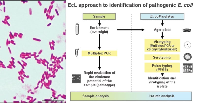
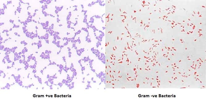
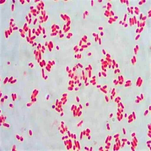
E coli is a Gramnegative rodshaped bacteria When Gram stained, the organism looks pink or red Here are a couple of pictures of a Gram stain of E coli that I did under the 100X objective lens on a standard light microscope You can of courseMODULE Morphology and Classification of Bacteria Microbiology 4 Notes 132 Microscopy The morphological study of bacteria requires the use of microscopes Microscopy has come a long way since Leeuwenhoek first observed bacteria using handground lenses The types of microscope are (i) Light or optical microscope (ii) Phase contrast microscope2 Micrococcus luteus or Staphylococcus epidermidis a Fix a smear of either Micrococcus luteus or Staphylococcus epidermidis to the slide as follows 1First place a small piece of tape at one end of the slide and label it with the name of the bacterium you will be placing on that slide 2Using the dropper bottle of deionized water found in the staining rack, place 1/2 of a normal sized

Solved 1 Identify The Morphology Morphological Arrangem Chegg Com
E coli morphology under microscope
E coli morphology under microscope-Escherichia coli bacteria on blood agar e coli under a microscope stock pictures, royaltyfree photos & images petri dishes with culture media for sarscov2 diagnostics, test coronavirus covid19, microbiological analysis e coli under a microscope stock pictures, royaltyfree photos & imagesD) Klebsiella & E) E Coli


Cell Shape And Cell Wall Organization In Gram Negative Bacteria Pnas
Most of the bacteria range from 022 µm in diameter The length can range from 110 µm for filamentous or rodshaped bacteria The most wellknown bacteria E coli, their average size is ~15 µm in diameter and 26 µm in length In this figure The size comparison between our hair (~ 60 µm) and E coli (~1 µm) Notice how small theIt is estimated that % of the human population are longterm carriers of S aureusAmong the most complex virions known, the T4 bacteriophage, which infects the Escherichia coli bacterium, has a tail structure that the virus uses to attach to host cells and a head structure that houses its DNA Adenovirus, a nonenveloped animal virus that causes respiratory illnesses in humans, uses glycoprotein spikes protruding from its
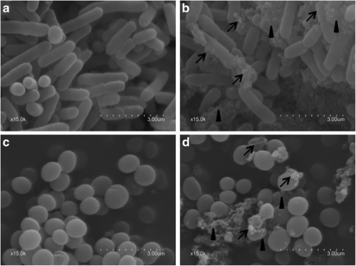
E coli under the microscope Escherichia coli (E coli) is a bacterium commonly found in various ecosystems like land and water Most of the strains of E coli are harmless, but some strains are known to cause diarrhea and even UTIs E coli is commonly studied as they are considered as a standard for the study of different bacteriaMost of the bacteria range from 022 µm in diameter The length can range from 110 µm for filamentous or rodshaped bacteria The most wellknown bacteria E coli, their average size is ~15 µm in diameter and 26 µm in length In this figure The size comparison between our hair (~ 60 µm) and E coli (~1 µm) Notice how small theWhat advantage does knowing t he morphology and arrangement of a culture provide Each bacterium is best treated with different antibiotics based on the cell's morphology and arrangement A physican can use this knowledge to diagnose the infectious organism and successfully prescribe a course of antibiotic therapy
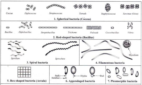
E faecalis can cause lifethreatening infections in humans, especially in the nosocomial (hospital) environmentEscherichia coli light microscopy Escherichia coli Magnification 1000×Unlike coccis bacteria, bacillus will appear as elongated rods (rodlike) when viewed under the microscope In most cases, the bacilli occur as single cells (eg Mycobacterium tuberculosis), but may occur in pairs (diplobacillus) or form chains commonly refered to as streptococcus (eg Bacillus cereus) Spirilla and V ibrio


Escherichia Coli E Coli An Overview Microbe Notes


Scanning Electron Microscopy The Morphology Of E Coli Cells Before Download Scientific Diagram
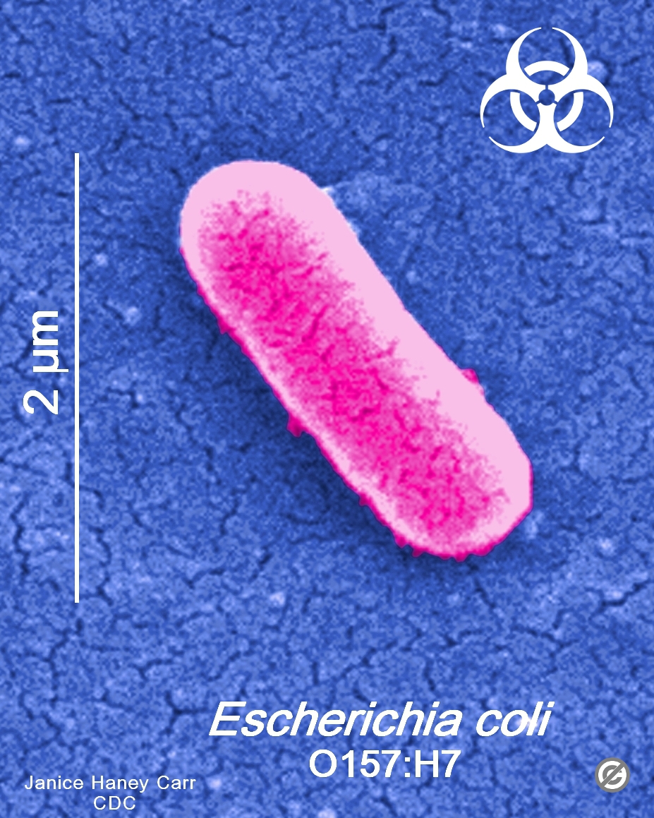
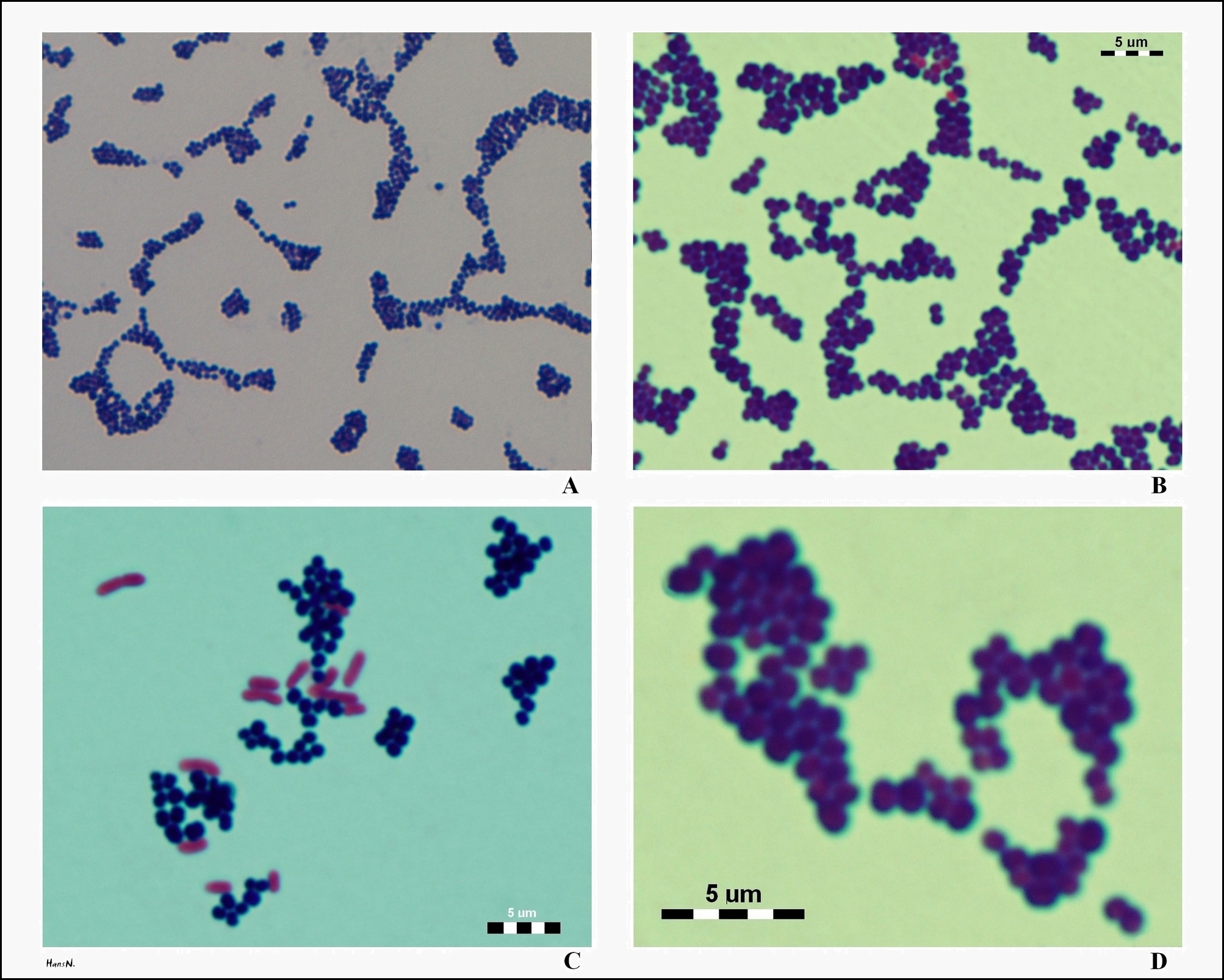
Escherichia coli (commonly abbreviated E coli) is a Gramnegative, rodshaped bacterium that is commonly found in the lower intestine of warmblooded organisms (endotherms) Most E coli strains are harmless, but some serotypes can cause serious food poisoning in humansWhere S is the Mueller matrix (), and θ and φ are polar and azimuth scattering angles, respectivelyThe operational angular range of the SFC was determined from analysis of polystyrene microspheres, as described in to be from 10° to 40°Optical Microscope Images of E coli cells were obtained with optical microscope Carl Zeiss Axio ImagerA1 using 100× oil immersion objective with 13Cocci in grapelike clusters (Saureus) and bacilli(Ecoli) Clinical significance of Saureus Frequently found as part of the normal skin flora on the skin and nasal passages;


Difference Between Scanning Electron Microscopy Sem And Transmission Electron Microscopy Tem Learn Microbiology Online
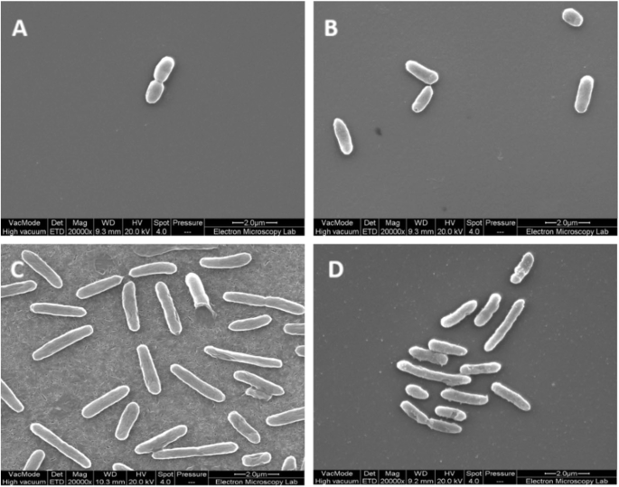


Thymol Tolerance In Escherichia Coli Induces Morphological Metabolic And Genetic Changes Bmc Microbiology Full Text
Use a dissecting/stereoscopic microscope for more detail Place the plate RIGHTSIDE UP on the stage, leaving the petri dish cover ON (Otherwise, your culture will become contaminated) There are 2 lenses on our scopes—10X and X the black lens knob is on the right side of the head of the microscopeEscherichia coli, often abbreviated E coli, are rodshaped bacteria that tend to occur individually and in large clumps E coli are classified as facultative anaerobes, which means that they grow best when oxygen is present but are able to switch to nonoxygendependent chemical processes in the absence of oxygenCell shape and E coli Depending on the growth conditions, E coli varies in size;


Staining Microscopic Specimens Microbiology
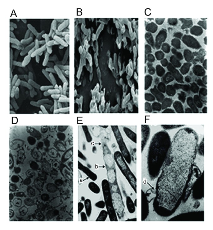


Enhancement Of Bacteriolysis Of Shuffled Phage Phix174 Gene E Springerlink
Nosepiece Holds 34 objectives2 Micrococcus luteus or Staphylococcus epidermidis a Fix a smear of either Micrococcus luteus or Staphylococcus epidermidis to the slide as follows 1First place a small piece of tape at one end of the slide and label it with the name of the bacterium you will be placing on that slide 2Using the dropper bottle of deionized water found in the staining rack, place 1/2 of a normal sizedGramstain Grampositive cocci Microscopic appearance Cocci in clusters, short chains, diplococci and single cocci Clinical significance Enterococcus faecalisis a Grampositive, commensal bacterium inhabiting the gastrointestinal tracts of humans and other mammals;


Gram Stain


Staining Microscopic Specimens Microbiology
It is routinely used as an initial procedure in the identification of an unknown bacterial species Let's suppose we have a smear containing mixture of Staphylococcus aureus and Escherichia coli as in previous case We will use the same stains as before and besides we will need Gram's iodine (strong iodine solution) and alcohol or acetoneAmong the most complex virions known, the T4 bacteriophage, which infects the Escherichia coli bacterium, has a tail structure that the virus uses to attach to host cells and a head structure that houses its DNA Adenovirus, a nonenveloped animal virus that causes respiratory illnesses in humans, uses glycoprotein spikes protruding from itsEntamoeba coli E coli cysts in concentrated wet mounts Cysts of Entamoeba coli are usually spherical but may be elongated and measure 10–35 µm Mature cysts typically have 8 nuclei but may have as many as 16 or more Entamoeba coli is the only Entamoeba species found in humans that has more than four nuclei in the cyst stage The nuclei may be seen in unstained as well as stained
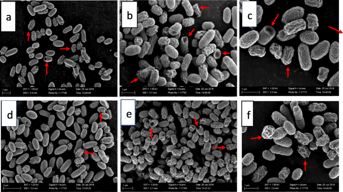


The Ultrastructural Damage Caused By Eugenia Zeyheri And Syzygium Legatii Acetone Leaf Extracts On Pathogenic Escherichia Coli Bmc Veterinary Research Full Text


Escherichia Coli On Macconkey Agar Colony Appearance Of Escherichia Coli Colony Morphology
We performed our study using E coli as the model organism, allowing us to place our results in the context of the relevant literature on E coli's physiology and cell size and growth rateInstead, their genetic material floats uncoveredGet help on Observing Bacteria and Blood Morphology and Other Microbiological Aspects on Graduateway Huge assortment of FREE essays & assignments The best writers!


Effect Of The Compound No On Cell Morphology Of E Coli Cells Download Scientific Diagram


Optical Microscope Images Of E Coli Cells Following Gram Staining A Download Scientific Diagram
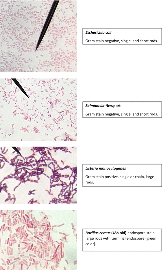
E coli is a Gramnegative rodshaped bacteria When Gram stained, the organism looks pink or red Here are a couple of pictures of a Gram stain of E coli that I did under the 100X objective lens on a standard light microscope You can of courseADVERTISEMENTS In this article we will discuss about 1 Definition of Bacteria 2 Morphology of Bacteria 3 General Methods of Classification 4 Nutrition, Respiration and Reproduction 5 Staining 6 Biochemical Test Contents Definition of Bacteria Morphology of Bacteria General Methods of Classifying Bacteria Nutrition, Respiration and Reproduction in Bacterial Cell Staining of BacteriaSUMMARY The shape of Escherichia coli is strikingly simple compared to those of higher eukaryotes In fact, the end result of E coli morphogenesis is a cylindrical tube with hemispherical caps It is argued that physical principles affect biological forms In this view, genes code for products that contribute to the production of suitable structures for physical factors to act upon
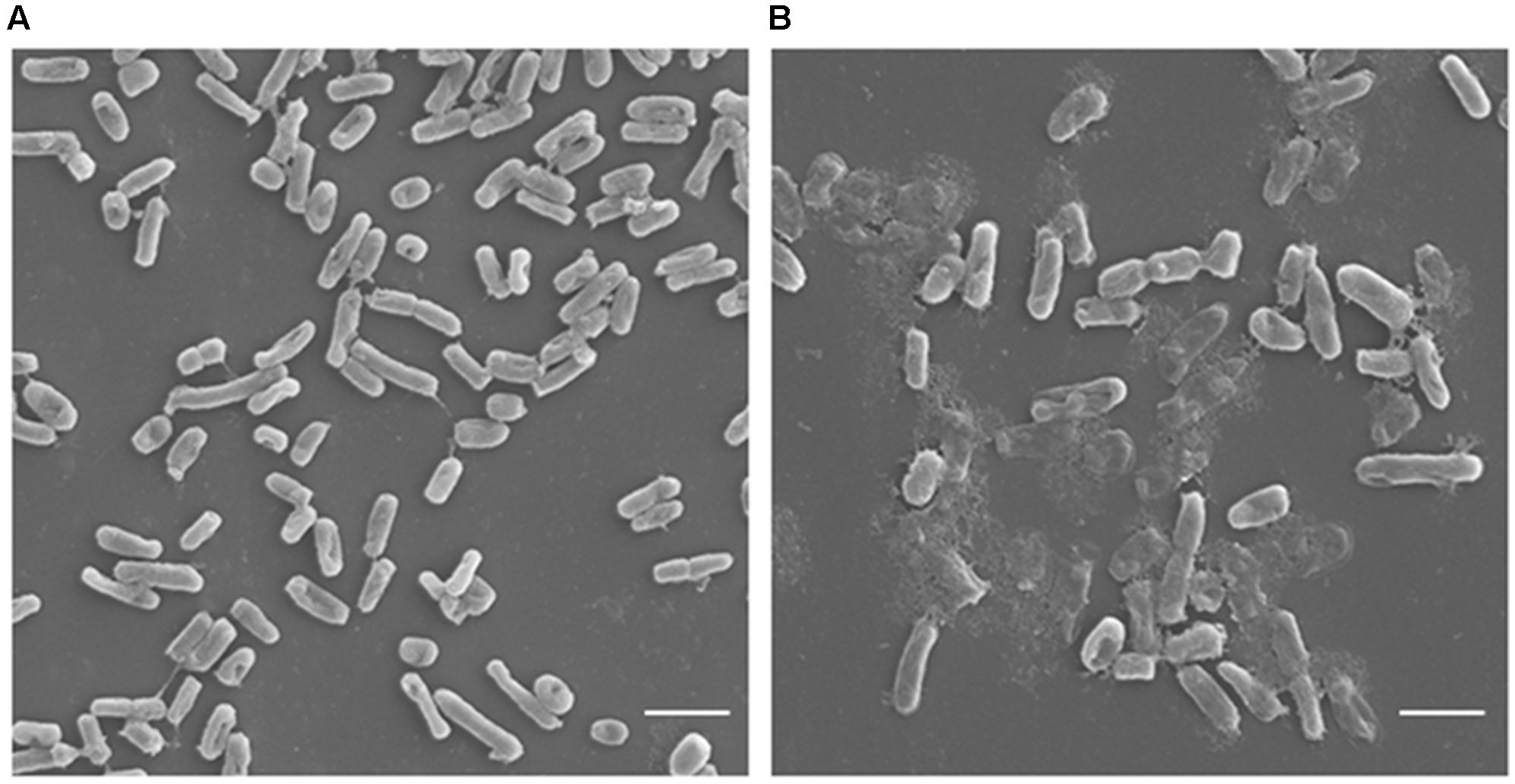


Frontiers Antimicrobial Potential Of Carvacrol Against Uropathogenic Escherichia Coli Via Membrane Disruption Depolarization And Reactive Oxygen Species Generation Microbiology


Staining Microscopic Specimens Microbiology
The microscope used to determine bacterial cell morphology is A) Compound light microscope B) Dissecting microscope A) Compound light microscope Under the microscope, rods are observed randomly scattered on the field The grouping pattern is E) E Coli B) Corynebacterium;Examples of Bacteria Under the Microscope Escherichia coli Escherichia coli (Ecoli) is a common gramnegative bacterial species that is often one of the first ones to be observed by students Most strains of Ecoli are harmless to humans, but some are pathogens and are responsible for gastrointestinal infections They are a bacillus shapedIn liquid culture media like peptone water and Nutrient broth, uniform turbidity is produced which is further analyzed for the morphology (under the microscope), gram reaction, biochemical tests, and staphylococcus specific tests That's all about the Morphology & Cultural Characteristics of Staphylococcus aureus


Escherichia Coli Wikipedia


Gram Stain Of E Coli Bacterium A Gram Stain Of Shows Gramnegative Download Scientific Diagram
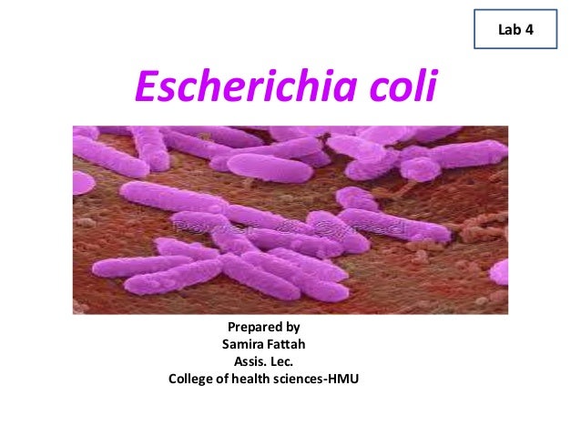
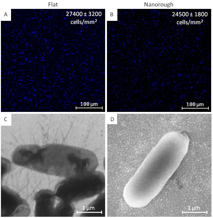
MORPHOLOGY OF ESCHERICHIA COLI (E COLI) Shape – Escherichia coli is a straight, rodIt is routinely used as an initial procedure in the identification of an unknown bacterial species Let's suppose we have a smear containing mixture of Staphylococcus aureus and Escherichia coli as in previous case We will use the same stains as before and besides we will need Gram's iodine (strong iodine solution) and alcohol or acetonePreparation of bacterial samples for morphology studies For each antibiotic concentration and each incubation time samples (2 ml) of each E coli strain were collected, washed three times with phosphatebuffered saline, and centrifuged The final pellet was divided, and each part was placed on a round microscope cover slide


Figure 2 From Morphological And Nanostructural Surface Changes In Escherichia Coli Over Time Monitored By Atomic Force Microscopy Semantic Scholar


12 3b Immediate Direct Examination Of Specimen Biology Libretexts
Allow students to explore the morphology of 1 of the most wellknown, Gramnegative bacteria, E coli This rodshaped, facultative anaerobe is normally present in the intestines of warmblooded animals However, it can be pathogenic This slide works well when discussing harmful and helpful bacteriaJane Buckle PhD, RN, in Clinical Aromatherapy (Third Edition), 15 VancomycinResistant Escherichia coli Escherichia is a gramnegative bacterium, which under the microscope is shaped like a rod with a small tail It is widely distributed in nature (Brooker 08)Escherichia coli (E coli) is part of the normal intestinal flora Some strains are pathogenic and can cause gastroenteritis, UTIEscherichia coli, often abbreviated E coli, are rodshaped bacteria that tend to occur individually and in large clumps E coli are classified as facultative anaerobes, which means that they grow best when oxygen is present but are able to switch to nonoxygendependent chemical processes in the absence of oxygen
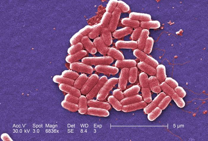


Details Public Health Image Library Phil


Pathogenic E Coli
Start studying Morphology of bacteria and gram stain Learn vocabulary, terms, and more with flashcards, games, and other study tools Ecoli Identify the bacteria (Gram ve Bacilli) Pseudomonas aeruginosa Connects eyepiece to microscope B?Morphology of E coli E coli is gramnegative (ve) rodshaped bacteria It is 13 x 0407 µm in size and 06 to 07 µm in volume It is arranged singly or in pairsPercentage of isolation of the five E coli Colonial Morphology (CM) classes from inpatients and outpatients Finally, through the electron microscope we carried out analysis of the colonies,


Biophysical Properties Of Escherichia Coli Cytoplasm In Stationary Phase By Superresolution Fluorescence Microscopy Mbio
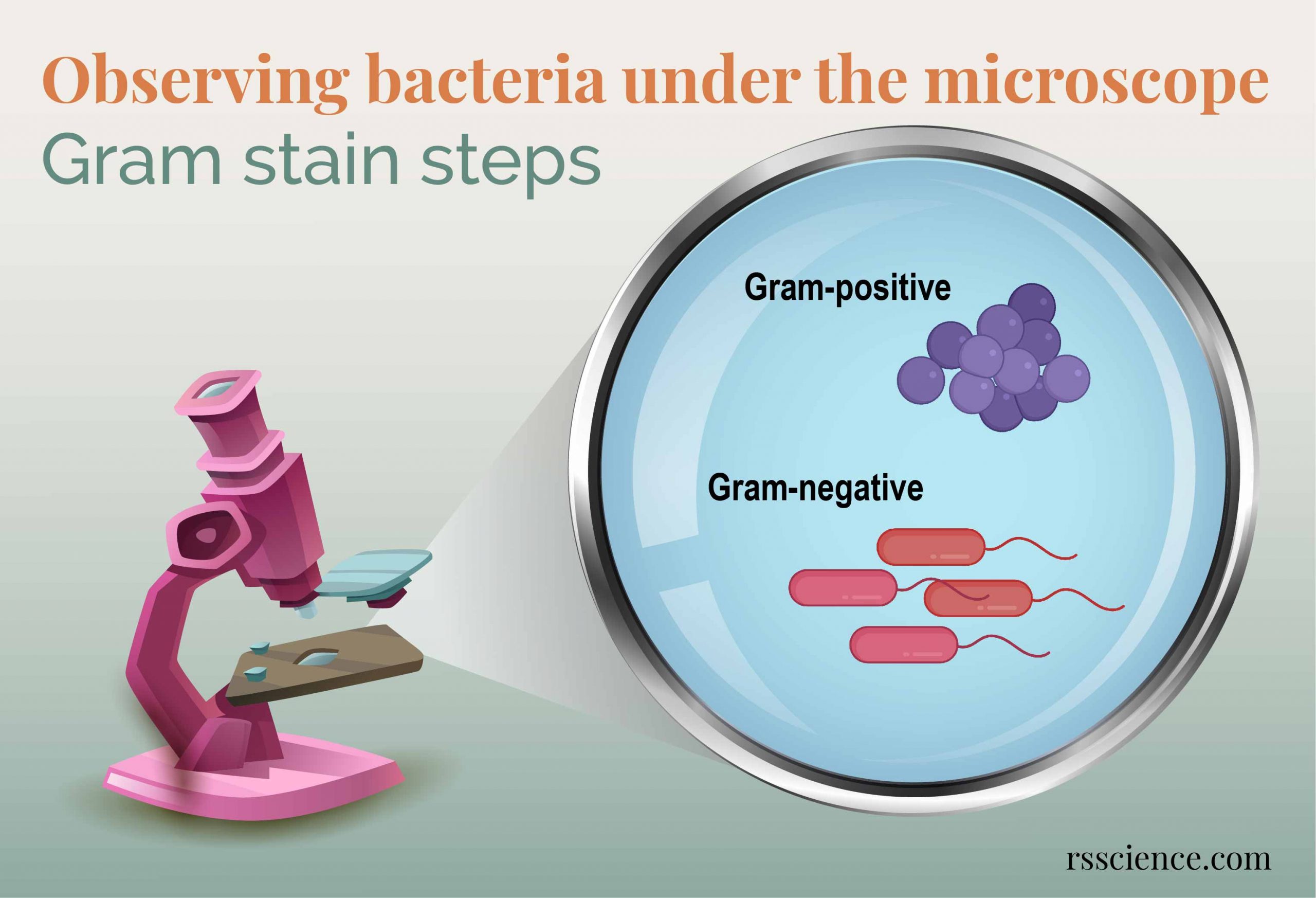


Observing Bacteria Under The Microscope Gram Stain Steps Rs Science
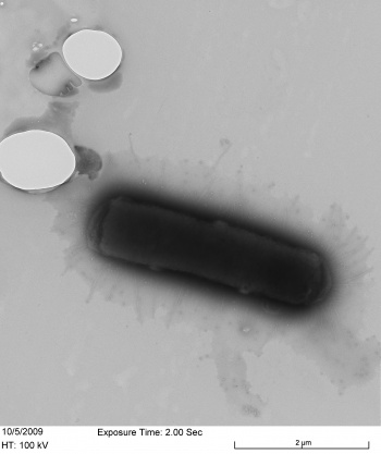
When viewed under the microscope, Gramnegative E Coli will appear pink in color The absence of this (of purple color) is indicative of Grampositive bacteria and the absence of Gramnegative E Coli Escherichia coli under 10х90х magnification using fuchsine as a dye by ElNokko (Own work) CC BYSA 40 (http//creativecommonsorg/licenses/bysa/40), via Wikimedia CommonsEntamoeba coli is a commensal of large intestine, but doesn't invade tissues Definitive diagnosis of amoebiasis depends on the demonstration of E histolytica trophozoite or cyst in stool Cases might get misdiagnosed because of similarities in their structure Following are the structural differences between E histolytica and E coliType and morphology E coli is a Gramnegative, facultative anaerobe (that makes ATP by aerobic respiration if oxygen is present, but is capable of switching to fermentation or anaerobic respiration if oxygen is absent) and nonsporulating bacterium Cells are typically rodshaped, and are about μm long and 025–10 μm in diameter, with a cell volume of 06–07 μm 3


The Rcs Stress Response And Accessory Envelope Proteins Are Required For De Novo Generation Of Cell Shape In Escherichia Coli Journal Of Bacteriology


Multicellular Behavior Of Environmental Escherichia Coli Isolates Grown Under Nutrient Poor And Low Temperature Conditions Sciencedirect
Percentage of isolation of the five E coli Colonial Morphology (CM) classes from inpatients and outpatients Finally, through the electron microscope we carried out analysis of the colonies,Escherichia coli (or simply E coli) is one of the many groups of bacteria that live in the intestines of healthy humans and most warmblooded animals E coli bacteria help maintain the balance of normal intestinal flora (bacteria) against harmful bacteria and synthesize or produce some vitamins However, there are hundreds of types or strainsFastgrowing cells are bigger than slowly growing ones (for a review, see reference123) At all growth rates, its shape can be roughly approximated by a cylinder with hemispherical ends


Pathogenic E Coli


Solved 1 Sketch The Four Way Streak For Isolation Your Chegg Com
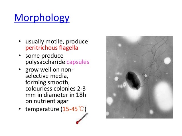
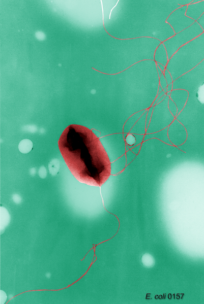
2 Morphology and Staining of Escherichia Coli E coli is Gramnegative straight rod, 13 µ x 0407 µ, arranged singly or in pairs (Fig 281) It is motile by peritrichous flagellae, though some strains are nonmotile Spores are not formed Capsules and fimbriae are found in some strains 3 Cultural Characteristics of Escherichia ColiEscherichia coli (E coli) is a large, varied group of bacteria found in the environment, foods and lower intestines of humans and animals( Sciencing,18) When observed under the microscope the shape identified was that of a spindle or rod shape2 Morphology and Staining of Escherichia Coli E coli is Gramnegative straight rod, 13 µ x 0407 µ, arranged singly or in pairs (Fig 281) It is motile by peritrichous flagellae, though some strains are nonmotile Spores are not formed Capsules and fimbriae are found in some strains 3 Cultural Characteristics of Escherichia Coli
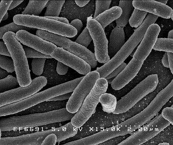


Escherichia Coli Microbewiki


E Coli
Biology Of E Coli E coli (Escherichia coli) are a small, Gramnegative species of bacteriaMost strains of E coli are rodshaped and measure about μm long and 0210 μm in diameterThey typically have a cell volume of 0607 μm, most of which is filled by the cytoplasm Since it is a prokaryote, E coli don't have nuclei;2 Micrococcus luteus or Staphylococcus epidermidis a Fix a smear of either Micrococcus luteus or Staphylococcus epidermidis to the slide as follows 1First place a small piece of tape at one end of the slide and label it with the name of the bacterium you will be placing on that slide 2Using the dropper bottle of deionized water found in the staining rack, place 1/2 of a normal sizedUse a dissecting/stereoscopic microscope for more detail Place the plate RIGHTSIDE UP on the stage, leaving the petri dish cover ON (Otherwise, your culture will become contaminated) There are 2 lenses on our scopes—10X and X the black lens knob is on the right side of the head of the microscope


Morphologies Of Recombinant E Coli Cells As Observed By Phase Contrast Download Scientific Diagram


Solved 1 Identify The Morphology Morphological Arrangem Chegg Com
Entamoeba coli is a nonpathogenic species of Entamoeba that frequently exists as a commensal parasite in the human gastrointestinal tract E coli (not to be confused with the bacterium Escherichia coli) is important in medicine because it can be confused during microscopic examination of stained stool specimens with the pathogenic Entamoeba histolytica
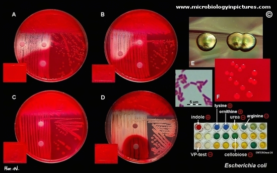


Escherichia Coli Colony Morphology And Microscopic Appearance Basic Characteristic And Tests For Identification Of E Coli Bacteria Images Of Escherichia Coli Antibiotic Treatment Of E Coli Infections
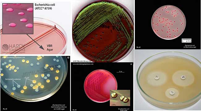


Escherichia Coli E Coli An Overview Microbe Notes


Www Mccc Edu Hilkerd Documents Bio1lab3 Exp 4 Pdf


Lab 1


Gram Stain Of E Coli Bacterium A Gram Stain Of Shows Gramnegative Download Scientific Diagram
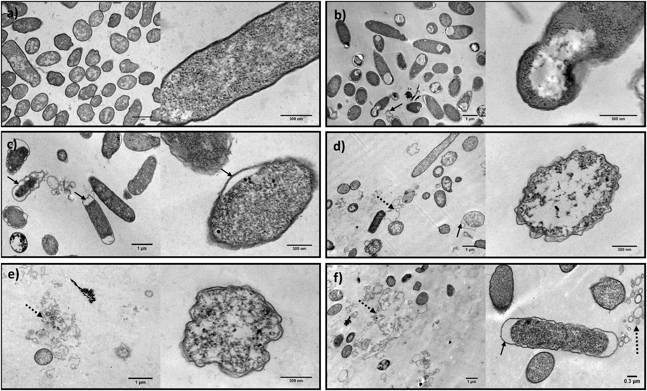


Frontiers Synergistic Antimicrobial Interaction Between Honey And Phage Against Escherichia Coli Biofilms Microbiology


Bacteria Under The Microscope E Coli And S Aureus Youtube


Gram Stain Wikipedia


Bacteria Capsules And Slime Layers Britannica


What Does An E Coli Bacteria Look Like Under A Microscope Quora


2 3 The Peptidoglycan Cell Wall Biology Libretexts


Usa Minnesota Firm Recalls Ground Beef Products Due To Possible E Coli O157 H7 Contamination Food Law Latest


Escherichia Coli Wikipedia


Catalysts Free Full Text Morphology Controlled Synthesis Of Zno Nanostructures For Caffeine Degradation And Escherichia Coli Inactivation In Water Html


Escherichia Coli Lecture


New Flat Embedding Method For Transmission Electron Microscopy Reveals An Unknown Mechanism Of Tetracycline Biorxiv


Toxins Free Full Text Identification Of A Novel Cathelicidin From The Deinagkistrodon Acutus Genome With Antibacterial Activity By Multiple Mechanisms Html


Thanatin Targets The Intermembrane Protein Complex Required For Lipopolysaccharide Transport In Escherichia Coli Science Advances


Escherichia Coli Colony Morphology And Microscopic Appearance Basic Characteristic And Tests For Identification Of E Coli Bacteria Images Of Escherichia Coli Antibiotic Treatment Of E Coli Infections


Enteric Bacteria List And Characteristics Medical Library


Staining Technology And Bright Field Microscope Use Springerlink


Gram Stain Images Microbiology Stain Study Tools


A Structural Study Of Escherichia Coli Cells Using An In Situ Liquid Chamber Tem Technology


Recombinant Production Of The Therapeutic Peptide Lunasin Microbial Cell Factories Full Text


Morphology Of E Coli Cells Under Microscope At 100 Magnification Download Scientific Diagram


Silver Coated Magnetic Nanocomposites Induce Growth Inhibition And Protein Changes In Foodborne Bacteria Scientific Reports


Escherichia Coli E Coli An Overview Microbe Notes


Escherichia Coli Wikipedia


Bioactivity And Morphological Changes Of Bacterial Cells After Exposure To 3 P Chlorophenyl Thio Citronellal Sciencedirect


Escherichia Coli Ecoliwiki


Introductory Chapter The Versatile Escherichia Coli Intechopen


Www Mccc Edu Hilkerd Documents Bio1lab3 Exp 4 Pdf


Molecular Response Of Escherichia Coli Adhering Onto Nanoscale Topography Nanoscale Research Letters Full Text


Cell Shape And Cell Wall Organization In Gram Negative Bacteria Pnas


Different Size Shape And Arrangement Of Bacterial Cells


Control Of Cell Shape In Bacteria Cell


Figure 3 From Influence Of Acetobacter Pasteurianus Sku1108 Asps Gene Expression On Escherichia Coli Morphology Semantic Scholar


Morphological And Physiological Changes Induced By High Hydrostatic Pressure In Exponential And Stationary Phase Cells Of Escherichia Coli Relationship With Cell Death Applied And Environmental Microbiology


S Aureus Under The Microscope Microscopic Appearance And Morphology Of S Aureus Cell Arrangement


Q Tbn And9gcqkye60ou Johpr02n Mbv1fferrjpdh Lnct7ymdf5qhyia1ld Usqp Cau
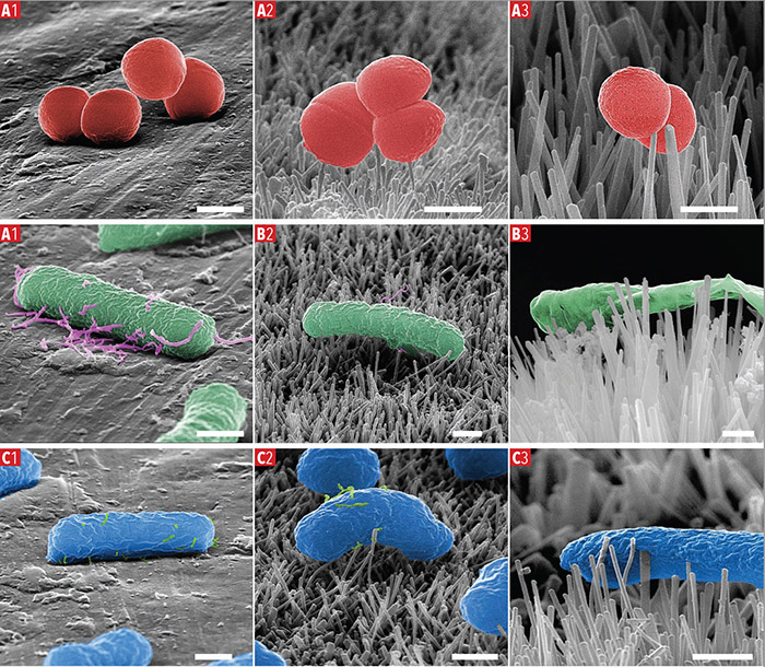


Characterisation Of Bactericidal Titanium Surfaces Using Electron Microscopy 18 Wiley Analytical Science


Escherichia Coli Light Microscopy


Filamentation By Escherichia Coli Subverts Innate Defenses During Urinary Tract Infection Pnas
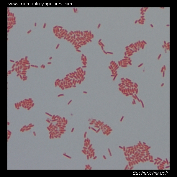


E Coli Gram Stain And Cell Morphology E Coli Micrograph Appearance Under The Microscope E Coli Cell Morphology E Coli Microscopic Picture
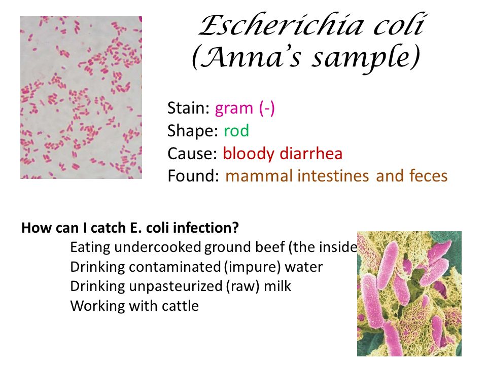


What Should You Have Seen Under The Microscope Activity Ppt Download


A Structural Study Of Escherichia Coli Cells Using An In Situ Liquid Chamber Tem Technology


Staphylococcus Aureus And Ecoli Under Microscope Microscopy Of Gram Positive Cocci And Gram Negative Bacilli Morphology And Microscopic Appearance Of Staphylococcus Aureus And E Coli S Aureus Gram Stain And Colony Morphology On Agar Clinical


Q Tbn And9gctgrmjg8acjrbxbniknzy1qewngtoobg8cixbwsv Ok9ny02ti0 Usqp Cau
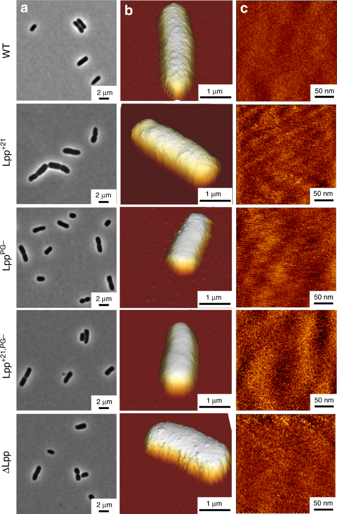


Lipoprotein Lpp Regulates The Mechanical Properties Of The E Coli Cell Envelope Nature Communications


Q Tbn And9gcqkye60ou Johpr02n Mbv1fferrjpdh Lnct7ymdf5qhyia1ld Usqp Cau


Gram Staining Principle Procedure Interpretation Examples And Animation


The Morphology Of Escherichia Coli Atcc And Staph Open I


Morphology Culture Characteristics Of Escherichia Coli E Coli


Escherichia Coli Slide W M Science Lab Microbiology Supplies Amazon Com Industrial Scientific


Bacteria And E Coli In Water


Gram Positive Bacteria Microbiology


Frontiers Phenotypic Changes Exhibited By E Coli Cultured In Space Microbiology


Representative Images Of Colony Coloration Microscopic Colony Morphology And Scattering Patterns Of E Coli Strains Grown On Each Of The Following Medium A Bhi Agar B Sorbitol Macconkey Agar Smac C Rainbow


Growth And Microscopic Morphology Of Recombinant E Coli S17 On Yt Download Scientific Diagram


Colony Characteristics Of E Coli Sciencing


Escherichia Coli Also Known As E Coli Ppt Video Online Download
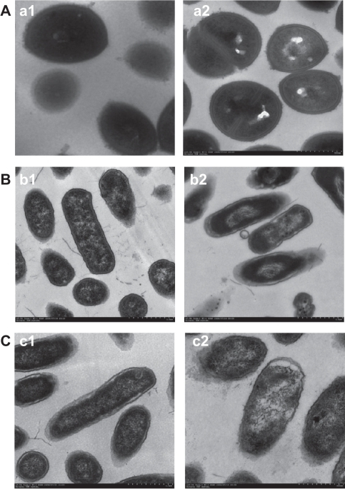


Morphology And Structure Of Bacterial Cells Under Trans Open I


Figure 1 From Bacteria Getting Into Shape Genetic Determinants Of E Coli Morphology Semantic Scholar


Antigen 43 From Escherichia Coli Induces Inter And Intraspecies Cell Aggregation And Changes In Colony Morphology Of Pseudomonas Fluorescens Journal Of Bacteriology


High Precision Characterization Of Individual E Coli Cell Morphology By Scanning Flow Cytometry Konokhova 13 Cytometry Part A Wiley Online Library


Q Tbn And9gcr1d8rkh2anc2byppttahm7ynyx Tlktllbaokmpm8efwa1tr0a Usqp Cau
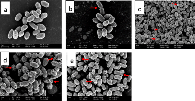


The Ultrastructural Damage Caused By Eugenia Zeyheri And Syzygium Legatii Acetone Leaf Extracts On Pathogenic Escherichia Coli Springerlink


Effects Of Sound Exposure On The Growth And Intracellular Macromolecular Synthesis Of E Coli K 12 Peerj
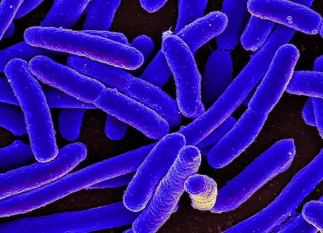


E Coli Under The Microscope Types Techniques Gram Stain Hanging Drop Method


Escherichia Coli E Coli Meaning Morphology And Characteristics


Staphylococcus Aureus And Ecoli Under Microscope Microscopy Of Gram Positive Cocci And Gram Negative Bacilli Morphology And Microscopic Appearance Of Staphylococcus Aureus And E Coli S Aureus Gram Stain And Colony Morphology On Agar Clinical
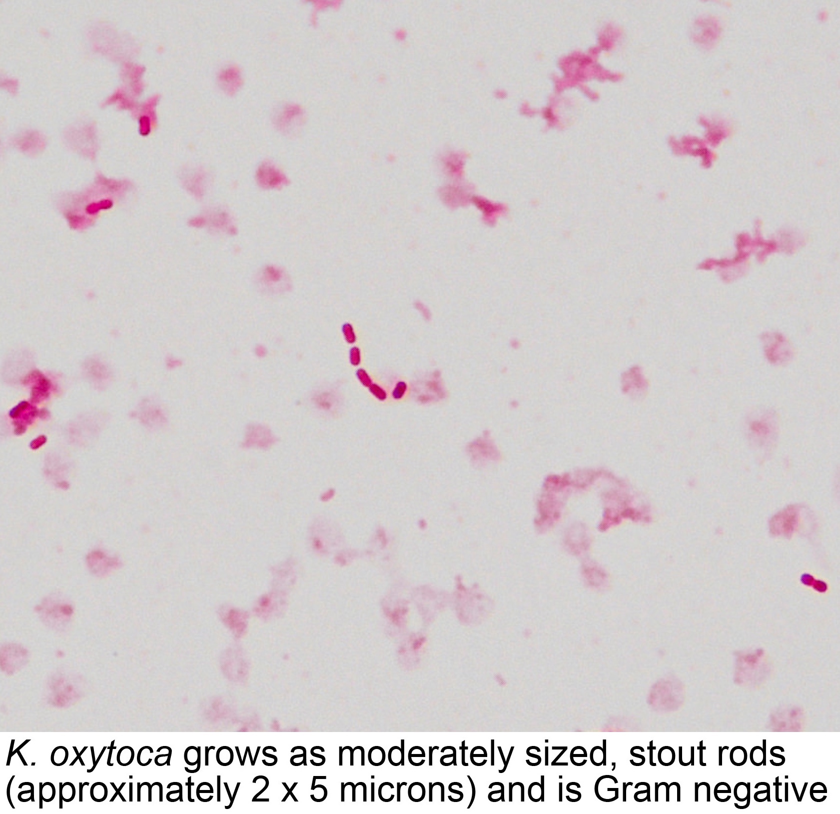


Pathology Outlines Klebsiella Oxytoca


Biol 230 Lab Manual Lab 1
/gram_positive_vs_negative-5b7f26d2c9e77c005746fbd7.jpg)


Gram Positive Vs Gram Negative Bacteria


コメント
コメントを投稿